Home

nøysom å tape bur mri brain scan labeled Autonomi Kamerat stropp

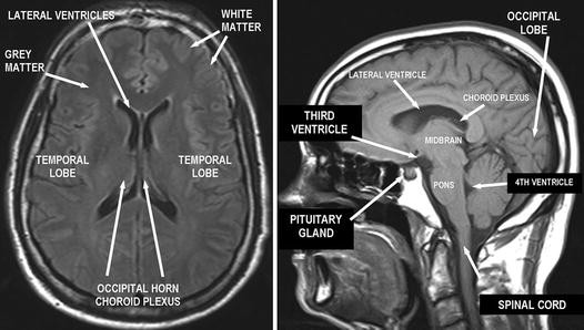
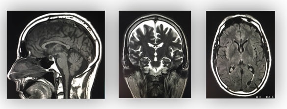
MRI anatomy | free MRI axial brain anatomy

A representative T1-weighted axial MRI of the retropalatal region in a... |  Download Scientific Diagram
A representative T1-weighted axial MRI of the retropalatal region in a... | Download Scientific Diagram

Labelled MRI of Normal Brain - Stock Image - C017/4418 - Science Photo  Library
Labelled MRI of Normal Brain - Stock Image - C017/4418 - Science Photo Library

A fast stochastic framework for automatic MR brain images segmentation

Ceditsa | Mri brain, Anatomy, Mri

Brain MRI: How to read MRI brain scan | Kenhub

MRI anatomy | free MRI axial brain anatomy

CNS Anatomy | SpringerLink

MRI Segmentation of the Human Brain: Challenges, Methods, and Applications

MRI anatomy | free MRI axial brain anatomy

9. Anterior-to-posterior sequence of coronal in situ magnetic resonance...  | Download Scientific Diagram
9. Anterior-to-posterior sequence of coronal in situ magnetic resonance... | Download Scientific Diagram

Frontiers | Longitudinal Evaluation of Cerebellar Signs of H-ABC  Tubulinopathy in a Patient and in the taiep Model | Neurology
Frontiers | Longitudinal Evaluation of Cerebellar Signs of H-ABC Tubulinopathy in a Patient and in the taiep Model | Neurology
NITRC: Manually Labeled MRI Brain Scan Database:

Comparative Overview of Brain Perfusion Imaging Techniques | Stroke

Mobile MRI brain atlas medical app review, NeuroRad Mini

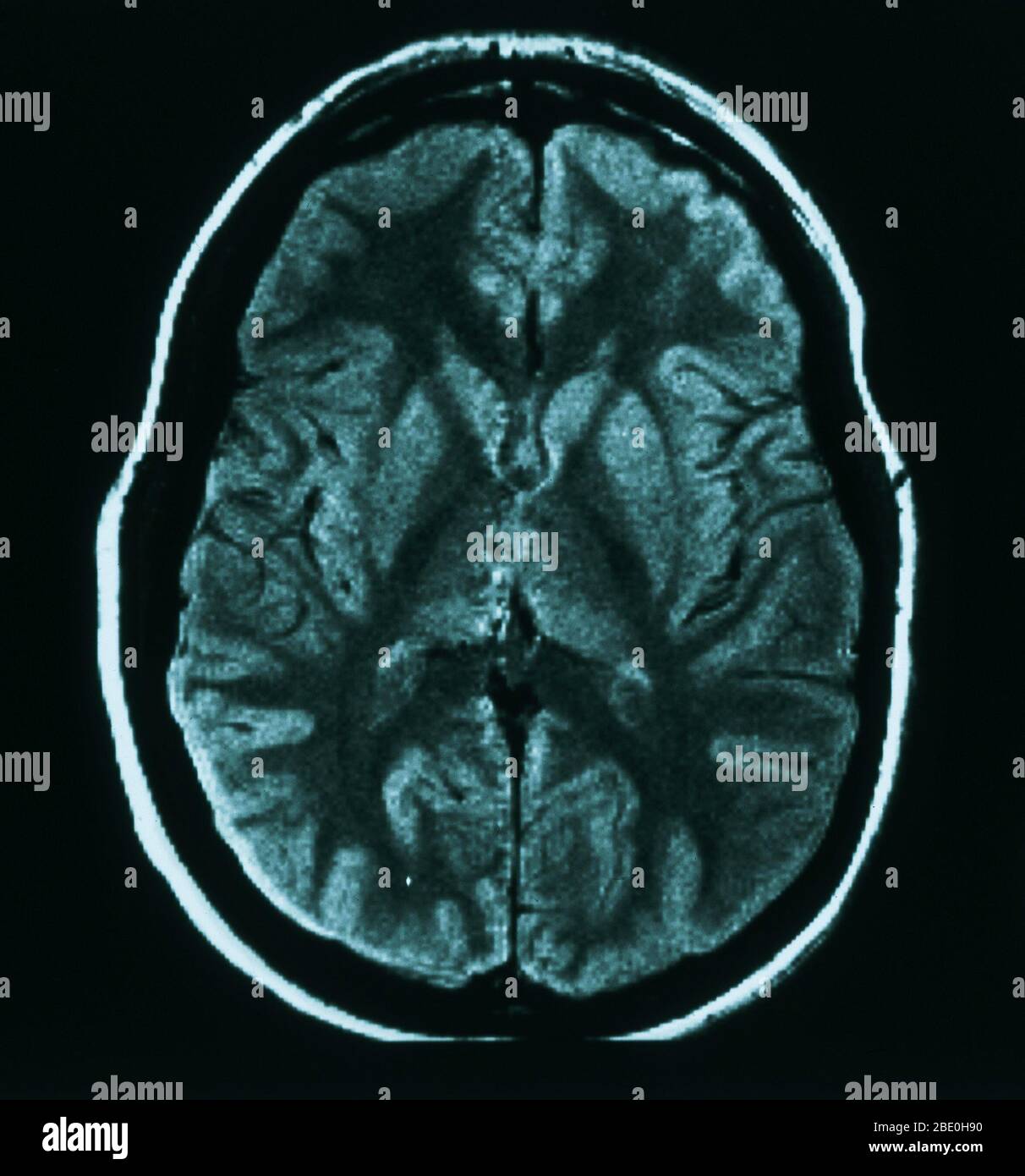
MRI scan (axial view) through the brain of a 31 year old female. The MRI is  normal Stock Photo - Alamy
MRI scan (axial view) through the brain of a 31 year old female. The MRI is normal Stock Photo - Alamy

Atlas | JHP MRI Brain Atlas

Brain lobes - annotated MRI | Radiology Case | Radiopaedia.org

MRI anatomy | free MRI axial brain anatomy

Cross-sectional anatomy of the brain - eAnatomy

Magnetic Resonance Imaging (MRI) Scanning - Principles - TeachMeAnatomy

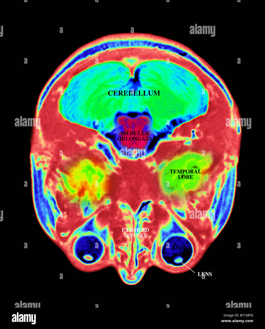
MRI scan of the brain, with the major structures labeled Stock Photo - Alamy

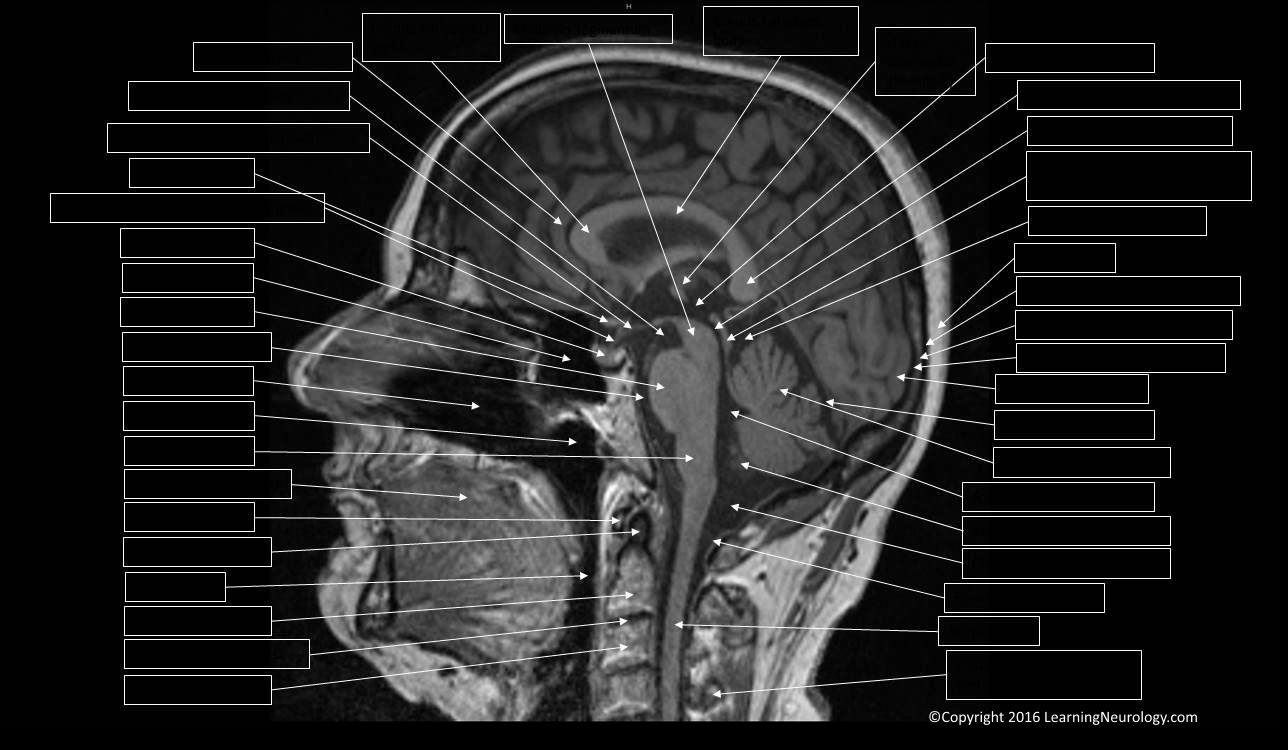
Approach to MRI brain | LearningNeurology.com

30 The brain MRI ideas | mri, brain, brain anatomy

Cross sectional anatomy of... - Radiology Students Of A.M.S | Facebook