Home

frem innblanding Summit d sign echo sti Slumber utarming

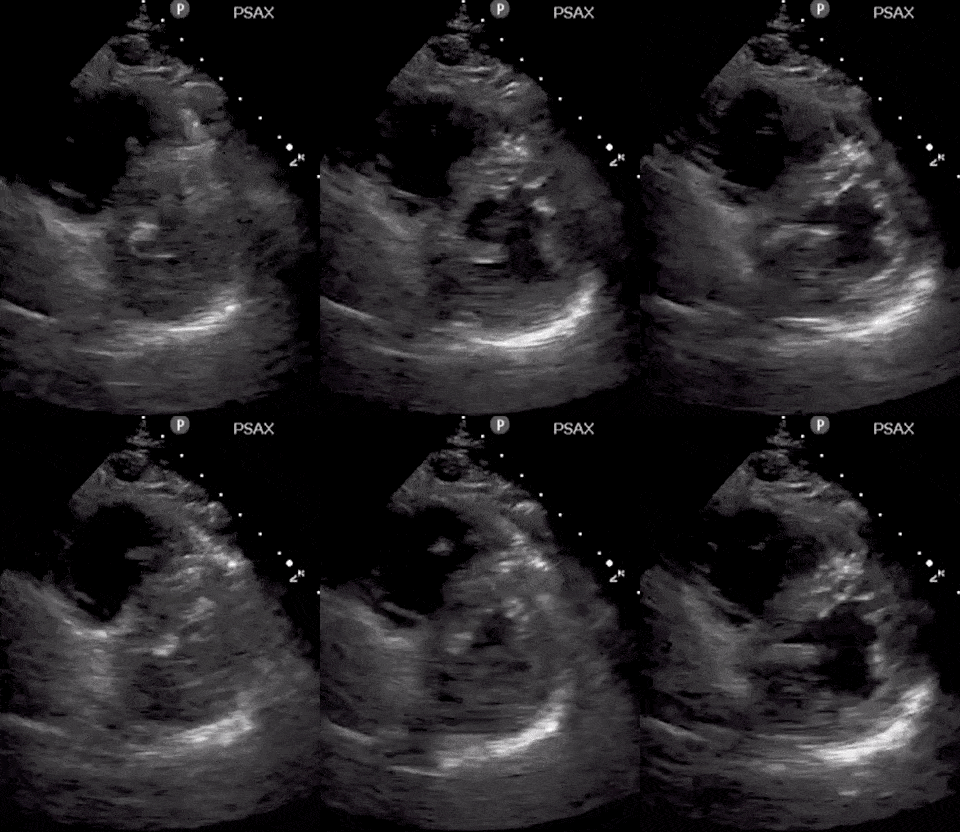
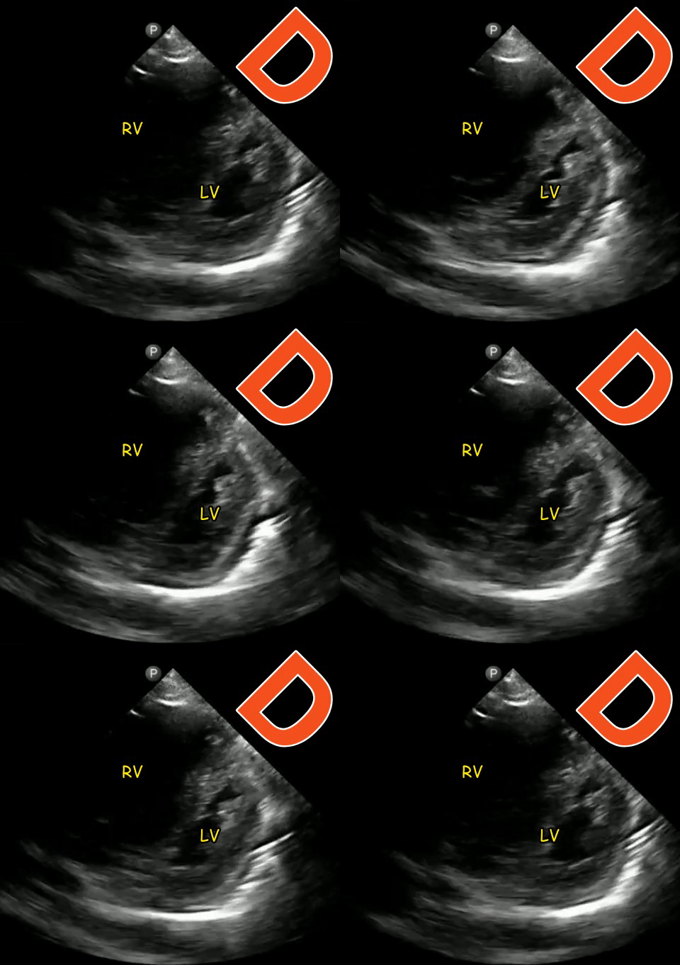
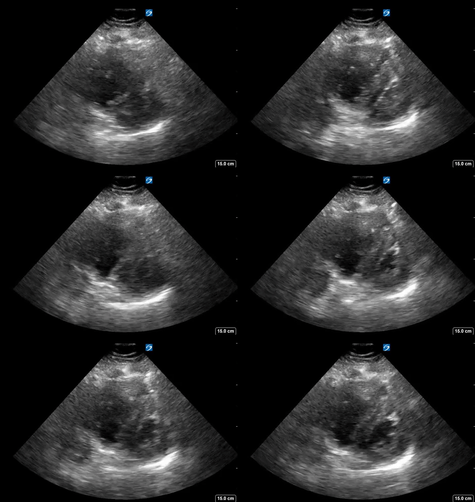
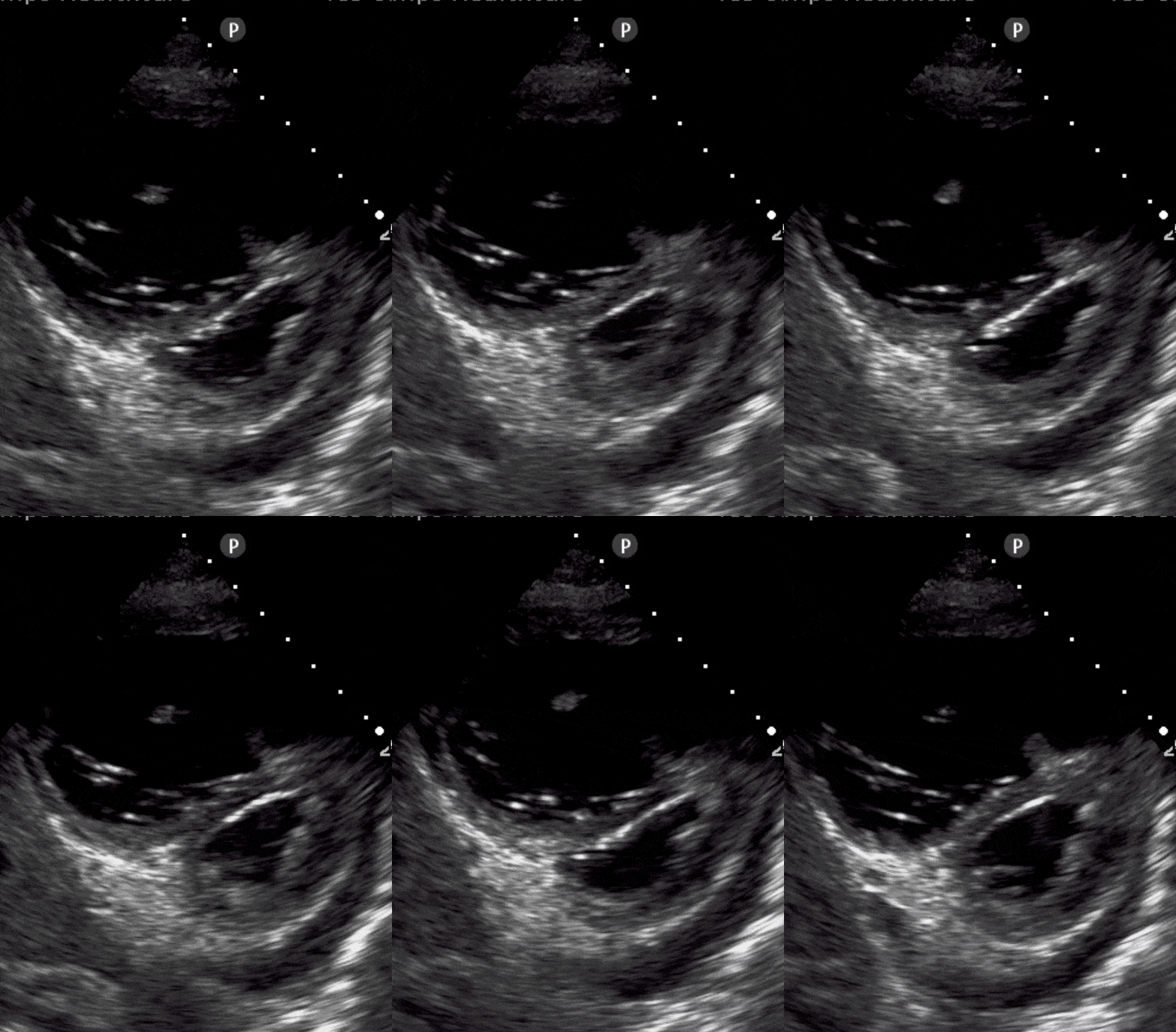
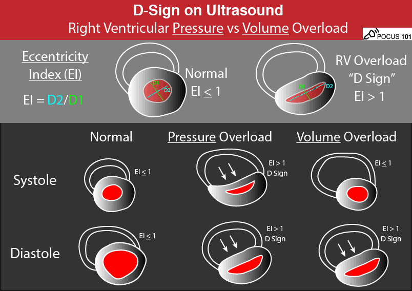
The D Sign - Right Heart Strain from Pressure vs Volume Overload - POCUS 101

The D Sign - Right Heart Strain from Pressure vs Volume Overload - POCUS 101

Everyday Ultrasound
Everyday Ultrasound

PDF] D-Shaped Left Ventricle, Anatomic, and Physiologic Implications |  Semantic Scholar
PDF] D-Shaped Left Ventricle, Anatomic, and Physiologic Implications | Semantic Scholar

D-shaped left ventricle. The interventricular septum is normally round... |  Download Scientific Diagram
D-shaped left ventricle. The interventricular septum is normally round... | Download Scientific Diagram

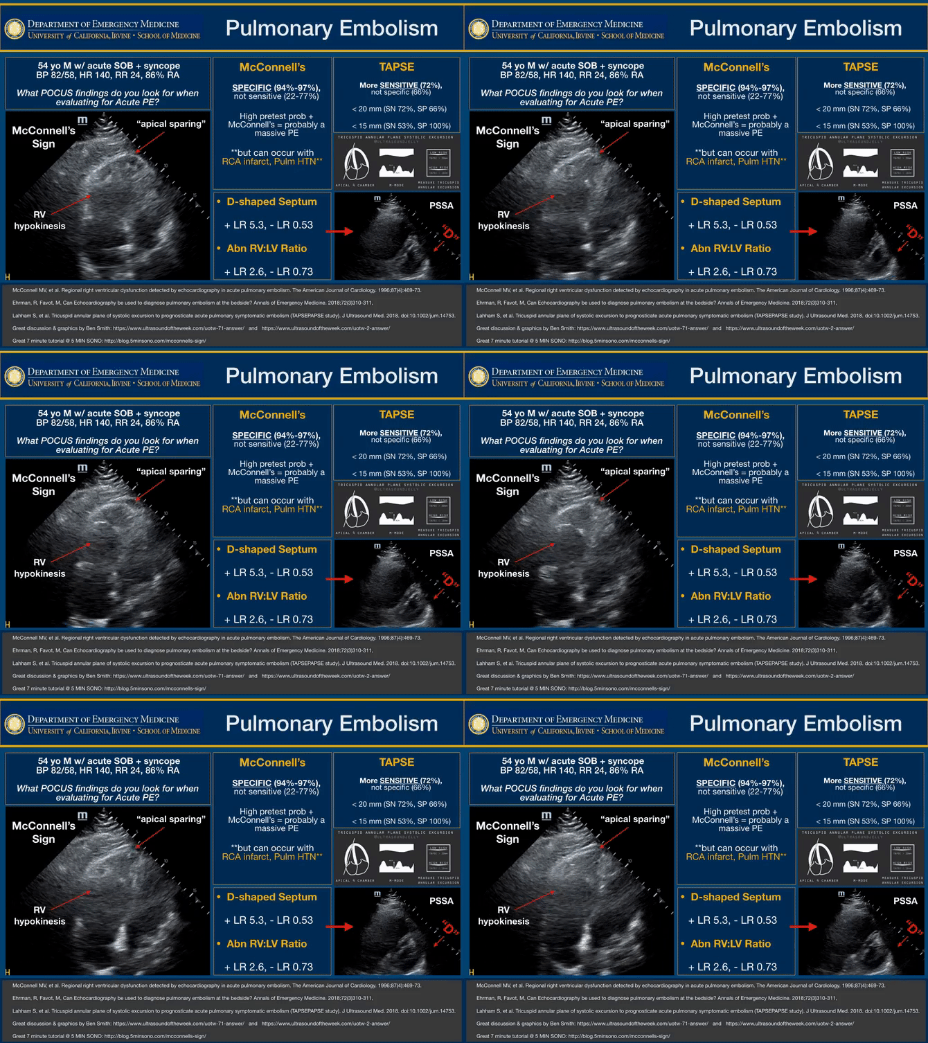
POCUS Findings in Acute Pulmonary Embolism - McConnell's ... | GrepMed

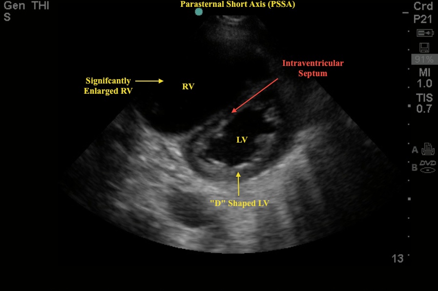
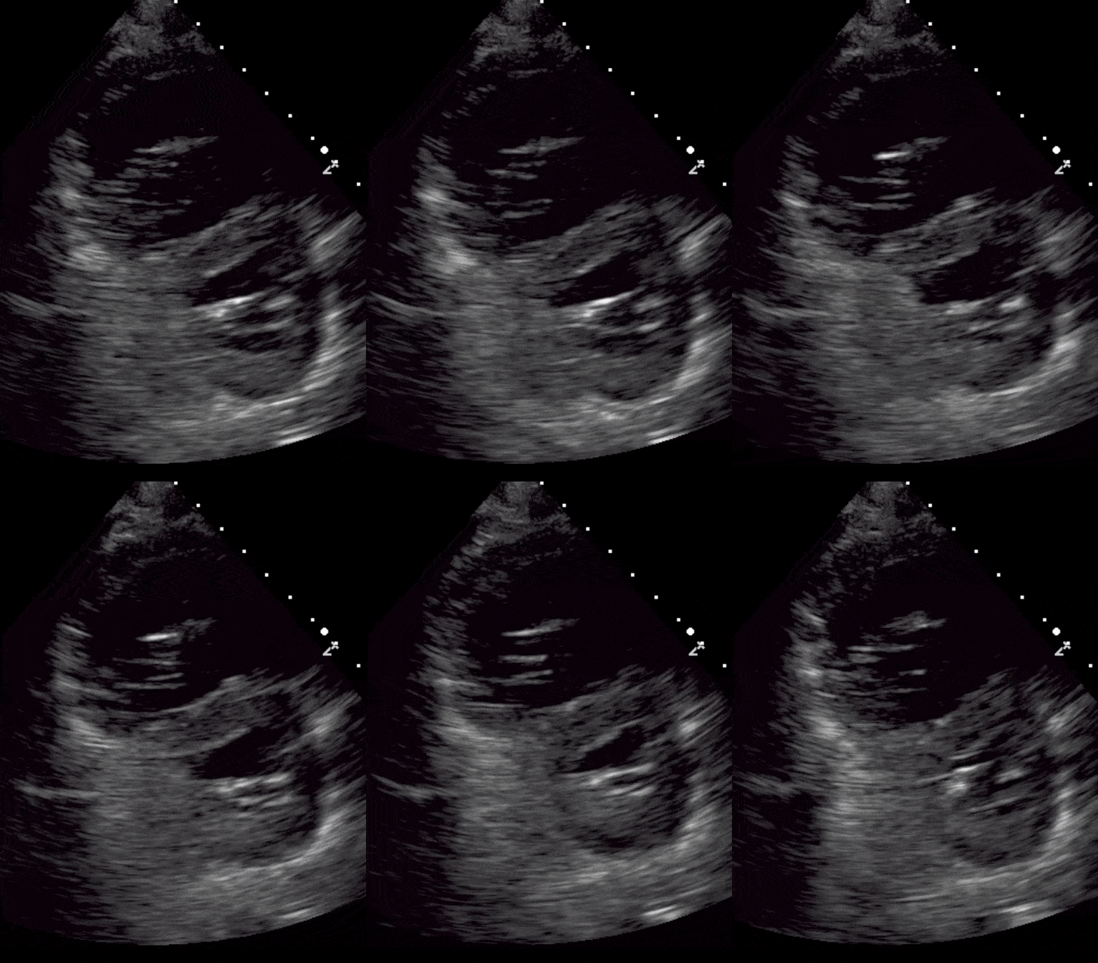
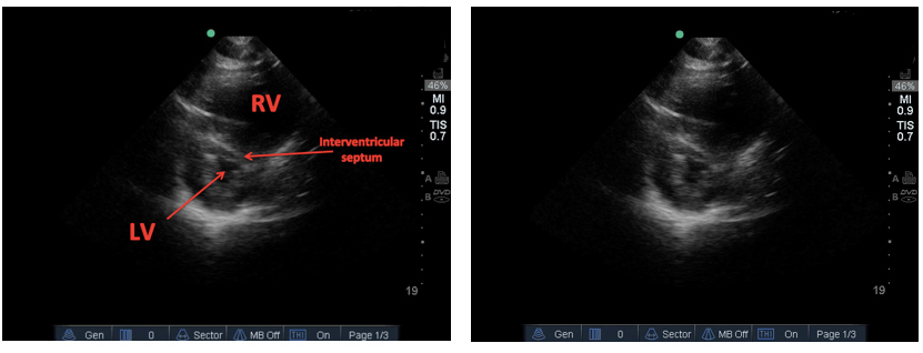
Focused Cardiac Ultrasound: Abnormalities on the Parasternal Short Axis  View - Renal Fellow Network
Focused Cardiac Ultrasound: Abnormalities on the Parasternal Short Axis View - Renal Fellow Network

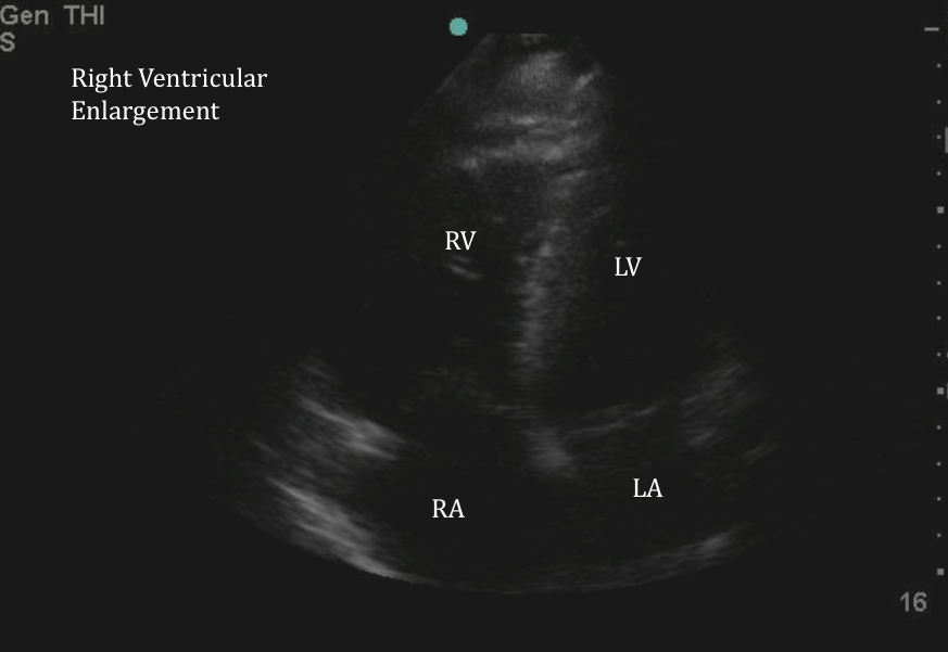
Enlarged RV | Emory School of Medicine

POCUS 101 on Twitter: "4) COVID-19 patients are hypercoagulable. Make sure  to look for Massive PE on echo: RV strain, D-sign, McConnell's sign, and  dilated IVC. https://t.co/RHBkyGgyBW https://t.co/ZHApdPVvRT" / Twitter
POCUS 101 on Twitter: "4) COVID-19 patients are hypercoagulable. Make sure to look for Massive PE on echo: RV strain, D-sign, McConnell's sign, and dilated IVC. https://t.co/RHBkyGgyBW https://t.co/ZHApdPVvRT" / Twitter

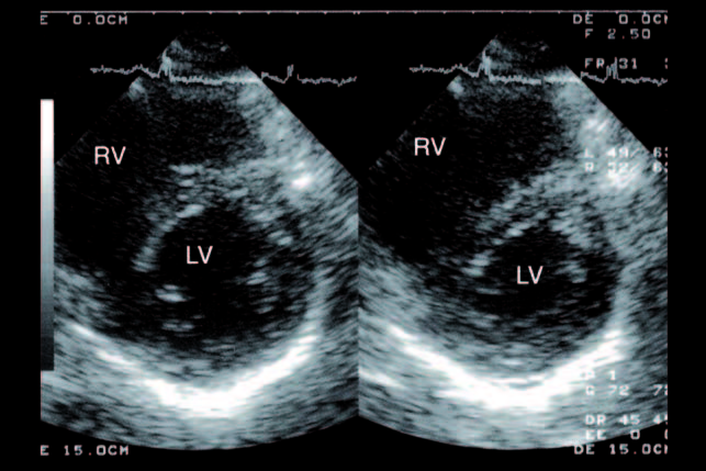
Transthoracic echocardiography, short-axis view (1A) and four-chamber... |  Download Scientific Diagram
Transthoracic echocardiography, short-axis view (1A) and four-chamber... | Download Scientific Diagram

Case of the Month #3 – Answer – Western Sono

Everyday Ultrasound
Everyday Ultrasound

The D Sign - Right Heart Strain from Pressure vs Volume Overload - POCUS 101

McConnell's Sign - a finding specific for Pulmonary Embolus | Emory School  of Medicine
McConnell's Sign - a finding specific for Pulmonary Embolus | Emory School of Medicine

The D Sign - Right Heart Strain from Pressure vs Volume Overload - POCUS 101

Hennepin Ultrasound | PEA Arrest: What's the Cause?

Right ventricular pressure overload- D-sign on Echo PSAX view - YouTube

Parasternal short axis view during systole in a patient with severe... |  Download Scientific Diagram
Parasternal short axis view during systole in a patient with severe... | Download Scientific Diagram

The D Sign - Right Heart Strain from Pressure vs Volume Overload - POCUS 101

McConnell's Sign - Point-of-Care Echo - YouTube

A Classic Pulmonary Embolism: Emergency Medicine Blog - Drexel University  College of Medicine
A Classic Pulmonary Embolism: Emergency Medicine Blog - Drexel University College of Medicine

D sign | thinking critical care

Echocardia - Wiki
Echocardia - Wiki

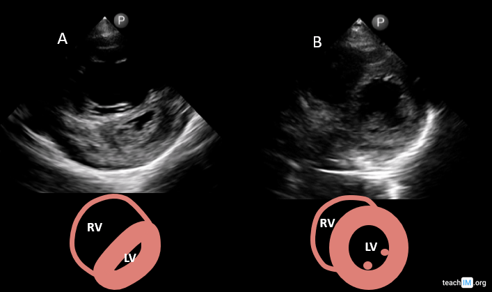
Right Ventricular (RV) Stain & "D-Sign" | POCUS | TeachIM